1. Quantitative biomedical imaging with nonlinear optical microscopy
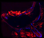
We employ multimodal nonlinear microscopy tools (SRS/CARS/SFG/TPEF) to quantitatively image biological tissues and structures. We are currently interested in the following topics:
2. Nonlinear optics of individual molecules and nanostructures
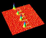
We are interested in characterizing the nonlinear optical properties of single nanostructures, including molecules. The use of plasmonics is an important aspect of this work. We are currently working on the following projects:
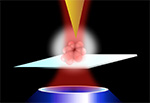
We combine the high resolution of scan probe techniques (~10 nm) with the molecular selectivity of nonlinear optical methods. A recent effort includes photo-induced force microscopy in the nonlinear optical regime. We currently focus on the following topics:
![]() Potma Labs, Department of Chemistry, Natural Sciences II, University of California, Irvine, CA 92697
Potma Labs, Department of Chemistry, Natural Sciences II, University of California, Irvine, CA 92697